Background and scenario
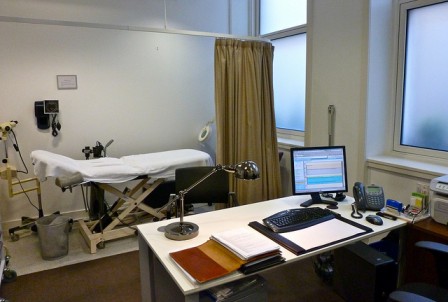
You’re a pathologist at a hospital and you’ve received a mislabelled tissue biopsy sample. Biopsies are invasive procedures and best not repeated for the patient’s sake. In order to best identify which tissue belongs to which patient biopsy, you’ll need to stain the tissue and make some observations. In order to complete your report, you’ll have to describe the cells in the tissue to the best of your ability by staining these cells with fluorophores, so you can visualize them under the microscope. These molecules can bind a variety of different structures on cells, including: actin, microtubules, DNA, ribosomes, and certain proteins associated with the mitochondria and ER. Part of preparing the samples involves placing these cells on a glass slide, treating them with your chosen fluorophores, and preserving them for prolonged storage. |
Map: Cells and tissues (2346)
|
||
|
Review your pathway |
