Examine other samples…
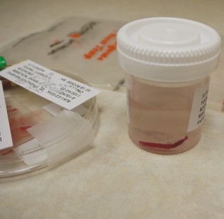
Uh oh. The freezer that held the labeling molecules broke down, so your stock of fluorophores is now very limited. On top of this headache, the rest of the actin-labeling molecules are unreliable, so you can no longer stain for ACTIN. For the rest of the samples, you'll only be able to stain for a maximum of THREE structures in order to ration what's left. Your goal is still to identify which biopsies belong to which patient based on your observations. Here is the information about the biopsies collected and from which patients: Alya Nirmala: A pap smear was performed (i.e. most superficial epithelial cells of the cervix were collected). These cells protect from abrasion and are organized as a stratified epithelium. Hye Su-Jin: Nasal polyps are suspected to interfere with Mr. Hye’s breathing. A biopsy was taken from his nasal cavity, where cells move mucous across their surface and are organized as a simple epithelium. Tao Lan: A biopsy was taken from his kidney tubules. These cells are absorptive and organized as a simple epithelium. Kenneth Dougal: Doctors suspect they have a rare disorder that causes low levels of leukocytes. A common way to confirm this diagnosis is by examining the morphology and organelles of the neutrophils, a multi-lobed and phagocytic white blood cell. Fatima Mubina: A biopsy was taken from her rectus femoris muscle (part of the quadricep muscle group), sectioned in cross-section, and stained. Doctors suspect Ms. Mubina has muscular dystrophy. Choose your next sample. |
Map: [For workshops and presentations] More cells and tissues (2492)
|
||
|
Review your pathway |
