Welcome!Who is in your group? List the names of students who are present.
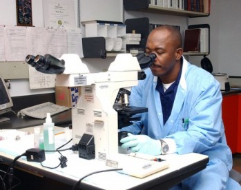
Pathologists are commonly asked to evaluate biopsied tissues for cancers and other diseases. Commonly, these healthcare professionals use histological stains to increase contrast in tissues that would otherwise be transparent. Other techniques involve fluorescence, which allows them to examine very specific structures in tissues. By using special molecules called fluorophores and special microscopes, pathologists can highlight specific parts of the cell to make their diagnoses. Briefly, fluorophores work by shining light of a certain wavelength onto the tissue specimen, stimulating the fluorophore to emit light at another wavelength, which is captured by the microscope. These fluorophores can be attached to molecules that bind specific structures in the cell, like actin, microtubules, DNA, and proteins specific to other organelles. Typically, there a limit to the number of fluorophores that can be used on a specimen at once, but as long as stocks aren't limited, pathologists can treat a specimen and observe separate structures in a tissue by preparing copies of the same specimen. |
Map: Cells and tissues [in class]_2 (2816)
|
||
|
Review your pathway |
